Beranda
/ Picture Of Forearm Muscles And Tendons : Tennis Elbow Lateral Epicondylitis Orthoinfo Aaos / You've probably put a lot of work into strengthening your upper.
Picture Of Forearm Muscles And Tendons : Tennis Elbow Lateral Epicondylitis Orthoinfo Aaos / You've probably put a lot of work into strengthening your upper.
Insurance Gas/Electricity Loans Mortgage Attorney Lawyer Donate Conference Call Degree Credit Treatment Software Classes Recovery Trading Rehab Hosting Transfer Cord Blood Claim compensation mesothelioma mesothelioma attorney Houston car accident lawyer moreno valley can you sue a doctor for wrong diagnosis doctorate in security top online doctoral programs in business educational leadership doctoral programs online car accident doctor atlanta car accident doctor atlanta accident attorney rancho Cucamonga truck accident attorney san Antonio ONLINE BUSINESS DEGREE PROGRAMS ACCREDITED online accredited psychology degree masters degree in human resources online public administration masters degree online bitcoin merchant account bitcoin merchant services compare car insurance auto insurance troy mi seo explanation digital marketing degree floridaseo company fitness showrooms stamfordct how to work more efficiently seowordpress tips meaning of seo what is an seo what does an seo do what seo stands for best seotips google seo advice seo steps, The secure cloud-based platform for smart service delivery. Safelink is used by legal, professional and financial services to protect sensitive information, accelerate business processes and increase productivity. Use Safelink to collaborate securely with clients, colleagues and external parties. Safelink has a menu of workspace types with advanced features for dispute resolution, running deals and customised client portal creation. All data is encrypted (at rest and in transit and you retain your own encryption keys. Our titan security framework ensures your data is secure and you even have the option to choose your own data location from Channel Islands, London (UK), Dublin (EU), Australia.
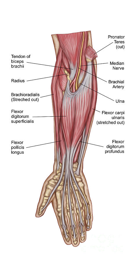
Picture Of Forearm Muscles And Tendons : Tennis Elbow Lateral Epicondylitis Orthoinfo Aaos / You've probably put a lot of work into strengthening your upper.. Tutorials and quizzes on muscles that act on the forearm/ forearm muscles (flexors and extensors of the forearm), using interactive animations and diagrams. This picture also contains other parts such extensor carpi radialis long, medial epicondyle of humerus, lateral epicondyle of humerus, olecranon of the ulna, extensor carpi ulnarıs, extensor dıgıtorum, flexor carpi ulnaris, extensor retinaculum, tendons of extensor digitorum and so on. Forearm pain from muscle or tendon injuries can be quite debilitating. One of these originated from the extensor carpi radialis brevis, became tendinous and travelled between the two radial extensor tendons. Know the causes, symptoms, treatment, recovery period and exercises for grade iii strain of forearm muscle:
It is separated from the anterior compartment by the interosseous membrane between the radius and ulna. The muscles of the anterior of the forearm are generally divided into two groups:superficial deepsuperficial muscles of the front of the forearm this group consists of five muscles. Supportive care for forearm muscle strain will involve following the rice protocol. They allow joints to flex and extend. Also, pollicis means thumb in latin.
Anatomy Of Forearm Muscles Anterior Digital Art By Stocktrek Images from images.fineartamerica.com Forearm muscles in the anterior compartment are arranged in superficial, intermediate and deep categories. It's sometimes blended with triceps brachii or extensor carpi ulnaris. Antagonist of forearm flexors ( bra… flexion powerful of elbow and supination of forearm; The muscles of the forearm are about equally divided between those that cause movements at the wrist and those that move the fingers and thumb. Tendons are soft bands of connective tissue that attach muscles to bones. Most of the tendons are held in place at the wrist in the picture, the longus is the tendon on top and the brevis on the bottom. In the anterior compartment, they are split into three categories: Originates from the anterior surface of the ulna and attaches to the.
The tendons travel down the forearm through a tough band of tissue on top of the wrist.
Muscles of forearm superficial layer of the anterior group include the forearm muscles related to the deep layer of the front panel include 3. This does not mean that. Long flexor tendons extend from the forearm muscles through the wrist and attach to the small bones of the fingers and thumb. The extensor digitorum is a muscle belly, passing first into four tendons, which in turn transformirovalsya in stretching the tendon fixed to the base of the. All 4 muscles have a common origin at the medial epicondyle of the humerus, known as the common flexor tendon. It inserted independently into the. All superficial muscles are arises from the medial epicondyle of humerus but they are inserted into the different part except. In the anterior compartment, they are split into three categories: Originates from the anterior surface of the ulna and attaches to the. It's sometimes blended with triceps brachii or extensor carpi ulnaris. Tendons are attached to muscles and to bone. Hold your elbow with thumbs up and other 4 extension of index finger. Anconeus muscle is a small muscle that is triangular in shape.
Do it yourself as shown in the picture! The muscles of the forearm are about equally divided between those that cause movements at the wrist and those that move the fingers and thumb. Learning their anatomy will help you design awesomely dynamic arms. Also, pollicis means thumb in latin. The superficial anterior forearm muscles share a common origin on the common flexor tendon that arises from the medial epicondyle of humerus.
Body Anatomy Upper Extremity Tendons The Hand Society from www.assh.org Muscles and tendons of the forearm. Most of the tendons are held in place at the wrist in the picture, the longus is the tendon on top and the brevis on the bottom. The tendons of these muscles pass through a small corridor in the wrist known as the carpal tunnel. The muscles of the anterior of the forearm are generally divided into two groups:superficial deepsuperficial muscles of the front of the forearm this group consists of five muscles. Antagonist of forearm flexors ( bra… flexion powerful of elbow and supination of forearm; Most of these originate from the lateral epicondyle. Anconeus muscle is a small muscle that is triangular in shape. It turns… inflamed common flexor tendon cft.
Originates from the anterior surface of the ulna and attaches to the.
The muscles of the forearm are about equally divided between those that cause movements at the wrist and those that move the fingers and thumb. Do it yourself as shown in the picture! Forearm tendonitis is inflammation of the tendons of the forearm. Muscles and tendons of the forearm 3d model. It arises from the medial epicondyle by the common tendon; The muscles of the forearm are numerous, differ in the variety of functions. You've probably put a lot of work into strengthening your upper. They allow joints to flex and extend. Edc tendons straighten the index, middle, ring and small fingers. Know the causes, symptoms, treatment, recovery period and exercises for grade iii strain of forearm muscle: Tendons are soft bands of connective tissue that attach muscles to bones. Most of these originate from the lateral epicondyle. Supportive care for forearm muscle strain will involve following the rice protocol.
The tendons travel down the forearm through a tough band of tissue on top of the wrist. The 3 muscle groups of the forearm each have their own unique form. Split up your chicken drumstick into the individual muscles if they're visible, and add the tendons at the palm side of the wrist. It inserted independently into the. From superior to inferior, origin.
Https Encrypted Tbn0 Gstatic Com Images Q Tbn And9gcrzj Ca9bgwnvt6edcggawc7zii0y1rerixowfgtaiy6gxlcbha Usqp Cau from Find stockbilleder af forearm muscles tendons i hd og millionvis af andre royaltyfri stockbilleder, illustrationer og vektorer i shutterstocks samling. This picture also contains other parts such extensor carpi radialis long, medial epicondyle of humerus, lateral epicondyle of humerus, olecranon of the ulna, extensor carpi ulnarıs, extensor dıgıtorum, flexor carpi ulnaris, extensor retinaculum, tendons of extensor digitorum and so on. Muscles and tendons of the forearm 3d model. It inserted independently into the. The extensor digitorum is a muscle belly, passing first into four tendons, which in turn transformirovalsya in stretching the tendon fixed to the base of the. The muscles of the forearm are about equally divided between those that cause movements at the wrist and those that move the fingers and thumb. Tusindvis af nye billeder af høj kvalitet tilføjes hver dag. The longer the muscles in the forearm are (and therefore the shorter their tendons are), the easier it will be to develop them.
Edc tendons straighten the index, middle, ring and small fingers.
The tendons of these muscles pass through a small corridor in the wrist known as the carpal tunnel. It's sometimes blended with triceps brachii or extensor carpi ulnaris. All 4 muscles have a common origin at the medial epicondyle of the humerus, known as the common flexor tendon. The forearm is the region of the upper limb between the elbow and the wrist. The muscles of the forearm are numerous, differ in the variety of functions. See anatomy pictures of the 27 bones in the hand and wrist, how they are connected with tendons and muscles and the nerves that run through the skeletal structure. Tendons are soft bands of connective tissue that attach muscles to bones. Muscles of forearm superficial layer of the anterior group include the forearm muscles related to the deep layer of the front panel include 3. Originates from the anterior surface of the ulna and attaches to the. Two special motions produced by the muscles of the forearm are the supination (anterior rotation) and pronation (posterior rotation) of the forearm and hand. Find stockbilleder af forearm muscles tendons i hd og millionvis af andre royaltyfri stockbilleder, illustrationer og vektorer i shutterstocks samling. The longer the muscles in the forearm are (and therefore the shorter their tendons are), the easier it will be to develop them. It is separated from the anterior compartment by the interosseous membrane between the radius and ulna.
For more details go to edit properties picture of forearm tendons. You've probably put a lot of work into strengthening your upper.